Tremor Analysis
Tremor in various parts of the body can occur as a symptom of different diseases. These include Parkinson's disease and essential tremor. Both belong to the most common neurological diseases. While Parkinson's disease has other symptoms such as slowed movements or rigid muscles, essential tremor is a description of the disease itself.
Distinguishing between different diseases based on tremor has proven to be difficult. The tremor characteristics of different diseases overlap and cannot be clearly distinguished. Therefore, misdiagnoses between different diseases is common. A therapeutic approach based on a misdiagnosis may lead to worsening of the symptoms and the disease progression.
Data from different types of sensors are used for this classification. Accelerometers are used to measure the acceleration of the hand tremor. Surface EMG electrodes are placed on the skin over the muscles under study to measure the action potential generated by different muscle fibers near the electrode. Both sensor types are available in the Trigno Avanti Sensor and the placement of the used sensors are shown below.
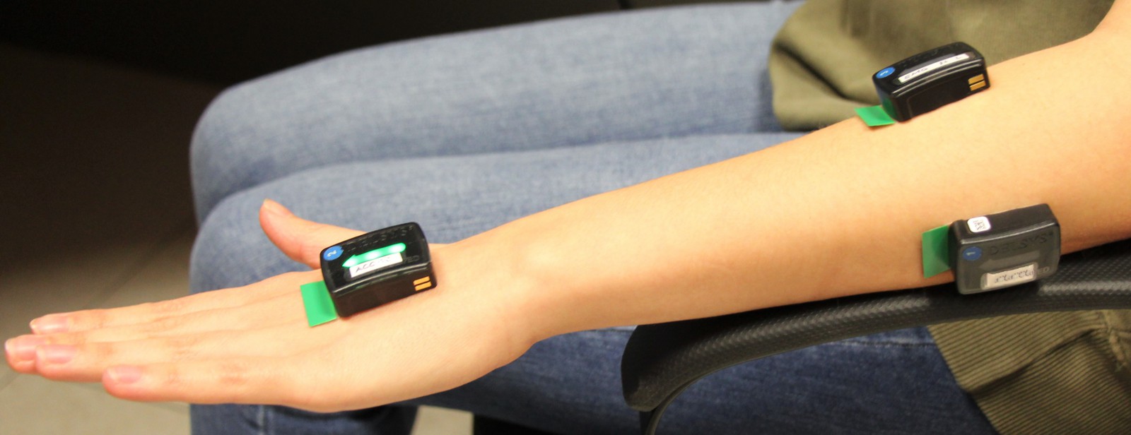
Trigno Avanti sensors.
Data acquired was used to develop the implemented signal processing chain. This processing chain can be seen in the main window of KiRAT under medical signal processing – neurologic signal processing – tremor analysis and is visualized in the screenshot below.
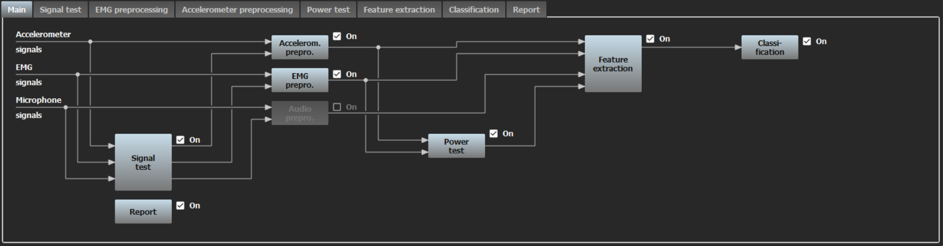
Screenshot of signal processing chain in KiRAT.
The signals are preprocessed subsequently. In tremor analysis, it is also possible to process an audio recording. Since this is not shown in this demo, the preprocessing of these signals is inactive. However, preprocessing only takes place if various signal tests are positive. For example, testing is done for a null signal or pure white noise. In a live demo with the Delsys system, the three axes of the acceleration sensor are combined into one signal. When data with only one axis is used, a trend removal takes place. The EMG signals are converted to a tremor signal via rectification. The power test also checks the signal quality. However, this tests the signals that have already been preprocessed.
These signals are used to extract various features and to classify the data. A decision tree and a simple neural network are used as classifiers. The signals, extracted features, and classification decision are combined into a report document.
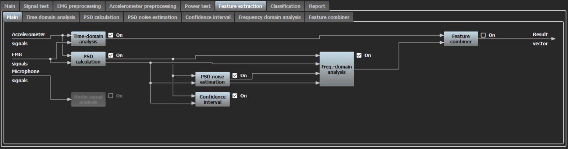
Screenshot of the feature extraction signal processing chain in KiRAT.
The feature extraction is divided into several modules, and the corresponding signal processing chain is shown above. In the time domain, features based on the amplitude of the signal are extracted. The amplitude, a side-by-side comparison of this and the change in this amplitude over time, is calculated. To extract features in the frequency domain, the power spectral density is calculated and a noise estimate is performed. Subsequently, features such as the peak frequency and the corresponding bandwidth can be calculated.
This demo can be used with a user interface that offers several options. In the picture below, a live examination with the Delsys system can be started. The current position is visualized and explained to the patient. In addition, the progress of the measurement is displayed. Meanwhile, a medical view can be used that shows different plots of the signals and the extracted features. Another way of using it is that already recorded data is processed and the results are summarized in a report. Individual measurements as well as many different patients can be evaluated.
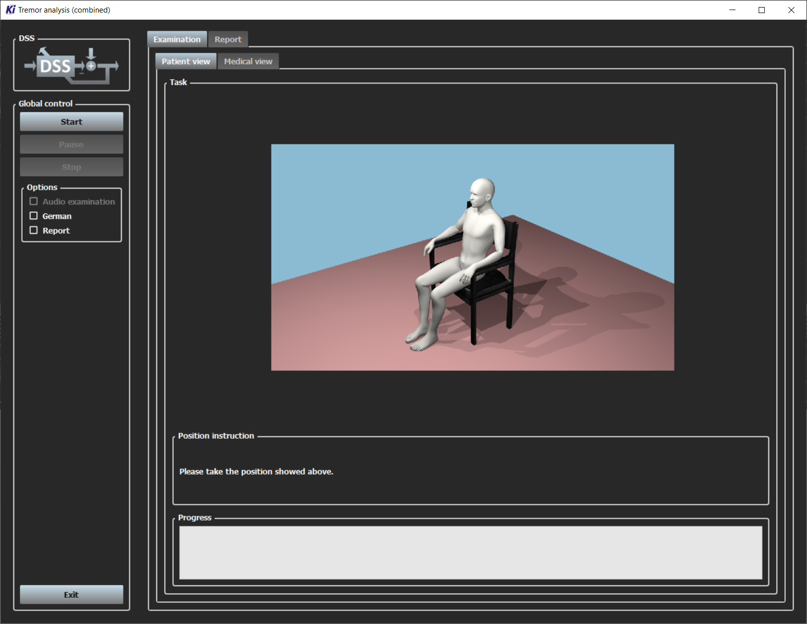
User interface of the tremor analysis.